Advanced Microscopy Facility
Welcome to the Advanced Microscopy Facility (AMF), an open user facility located in the Bob Wright Centre at the University of Victoria in beautiful British Columbia. We are one of the core labs of the Centre for Advanced Materials and Related Technology (CAMTEC).
What we are about
"To make it as easy as possible to visualize the invisible"
We offer high-end instrumentation and expertise in a world class facility, providing a welcoming and professional atmosphere. Our goal is to make it as easy as possible for our users to obtain scientific results of high quality.
Microscopy
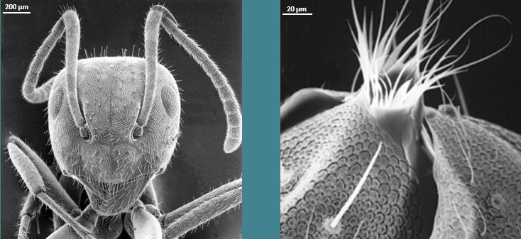
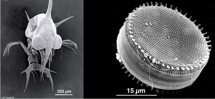
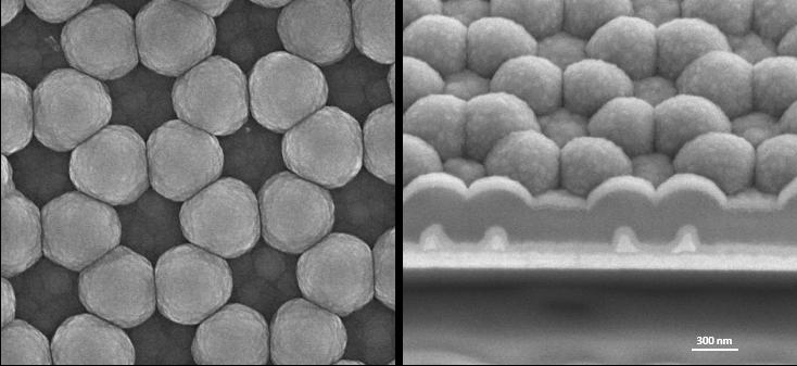
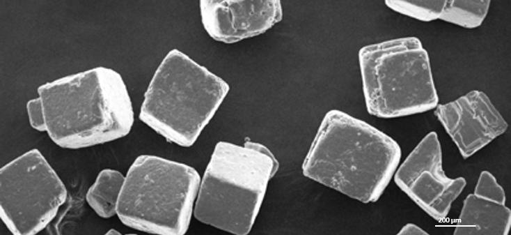
Microscopy is the science of obtaining magnified images of samples in order to reveal otherwise invisible details and better understand their morphology and structure. Microscopy includes also the techniques related to image interpretation, analysis and sample preparation (staining, fixing, structure reconstruction, etc.). Traditionally it is divided into two main areas: materials science and life science.
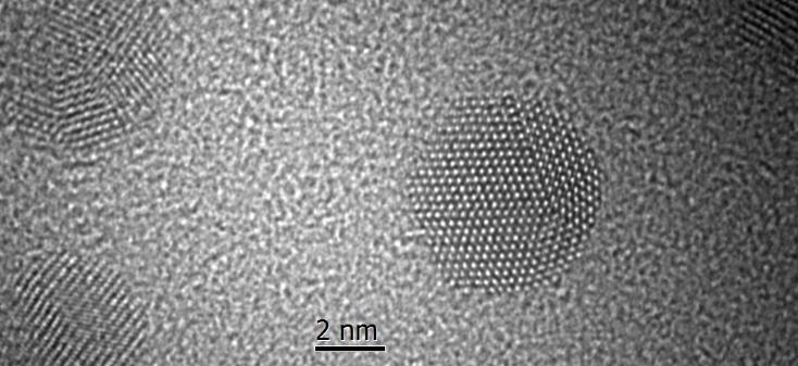
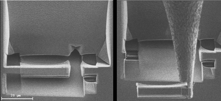
Information about the crystal structure of materials can be obtained by electron diffraction. Furthermore, instruments that measure the energy exchange between the illumination and the specimen or the resulting radiation emitted by the sample can provide information beyond simple imaging such as elemental composition or electronic structure. Examples of these techniques are Electron Energy Loss Spectroscopy (EELS) and Energy Dispersive X-ray Spectroscopy (EDXS).
Instruments
- STEM/TEM with aberration correction, EELS/EFTEM, EDX, holography and tomography.
- FIB with W deposition and nanomanipulator
- SEM with BSD and EDX
- TEM with digital imaging
- Sample preparation (C and Au/Pd coating, ion milling, UV and plasma cleaning, cryo-plunge)
Our People
![]()
- Co-Director: Rodney Herring
- Co-Director & HHTC Research Chair: Arthur Blackburn
- Lab Manager: Elaine Humphrey
- CAMTEC Manager: Milton Wang
- HHTC Engineer: Jonathan Rudge