Meet our graduates

Chelsea A.S. Dunning
Dr. Chelsea Dunning was interested in science and naturally got good grades as a high schooler, but struggled a lot with physics. Her parents had to force her to take 11th grade physics, which was a university entrance requirement! Her apprehension of physics was eased by how fundamental it was in understanding the world around us (and that it was math in disguise), and put in a lot of work to succeed in her physics courses. She grew to appreciate modern physics because it challenged her, and found fulfillment in learning the really cool stuff such as quantum mechanics, radiation, the Big Bang, and antimatter.
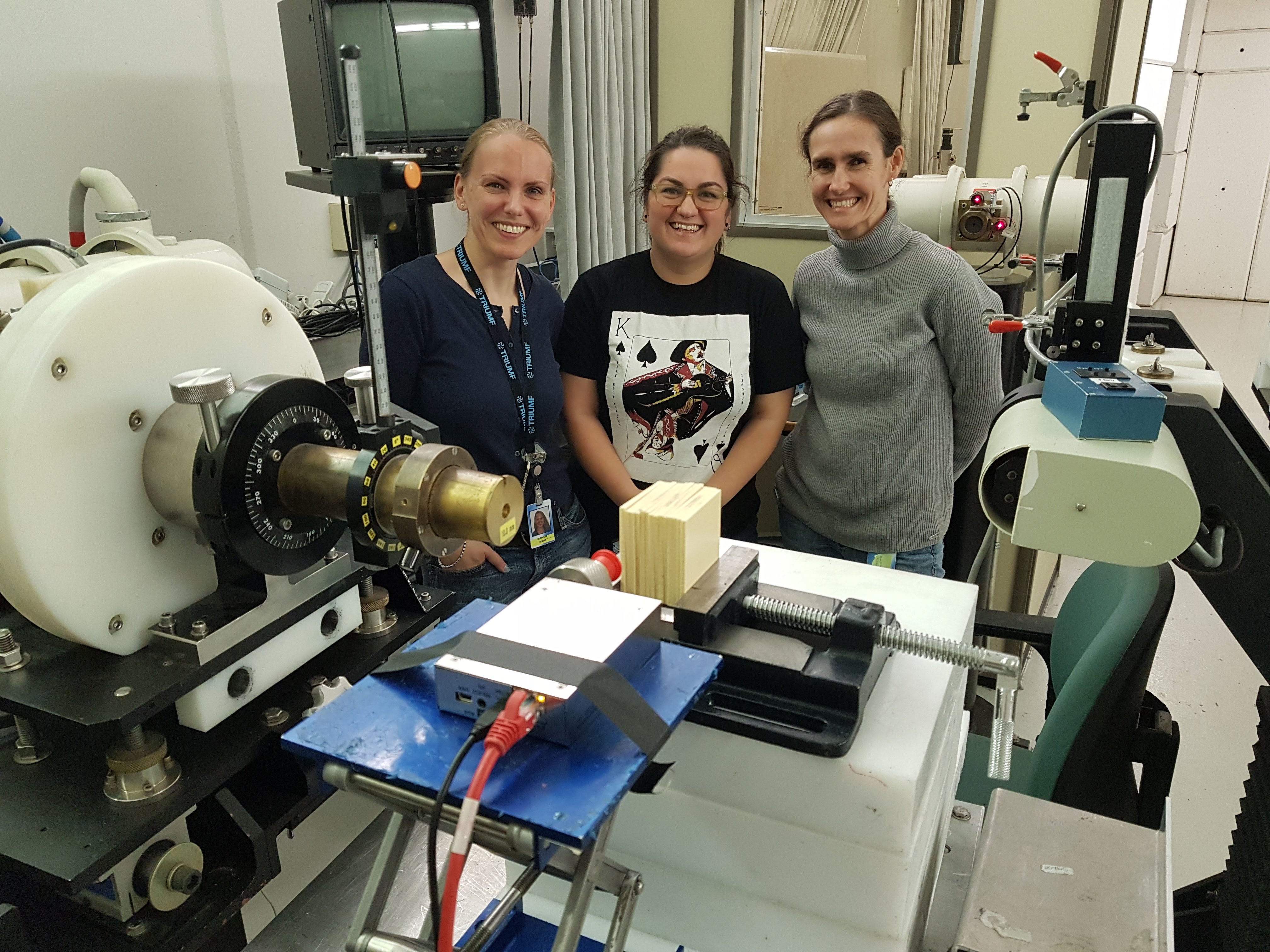
Chelsea obtained her Bachelor’s in Physics (Honours) at the University of British Columbia in Vancouver, BC, where she was fortunate enough to complete four co-op work terms at TRIUMF, Canada’s National Laboratory for Particle and Nuclear Physics. Her work experience ranged from health physics/radiation safety to detector prototype development, culminating in her undergraduate thesis work in proton eye therapy after completing her work terms. It was during her time at TRIUMF where she grew passionate about the role of physics in medicine, and her strong connections made at TRIUMF encouraged her to complete her PhD in Medical Physics at the University of Victoria.
At the University of Victoria, Chelsea was fortunate enough again to begin her Master’s degree at the same time her supervisor Dr. Magdalena Bazalova-Carter arrived as a professor. She really enjoyed working with Dr. Bazalova-Carter on Monte Carlo simulations of x-ray fluorescence computed tomography (XFCT) using her existing background on simulations as Dr. Bazalova-Carter established the XCITE lab. Her Monte Carlo work on XFCT, which investigated the impact of collimation geometries, spectrometer orientations, and excitation sources on XFCT image quality, led to three papers published in Medical Physics, Physics in Medicine & Biology, and IEEE Transactions on Medical Imaging, respectively. The first two papers each received the Best in Physics distinction at the 2018 and 2019 AAPM Annual Meetings in Nashville and San Antonio, United States, and she was recognized as a finalist in the Young Investigator Symposium at the 2017 COMP Annual Scientific Meeting in Ottawa for her work on collimation geometries in sheet beam XFCT. She enjoyed working so much with Dr. Bazalova-Carter, that she did the Master’s to PhD transfer, starting an apparent trend in the XCITE lab among Dr. Bazalova-Carter’s Master’s students :)
Chelsea really liked the flexibility of her work projects; she became interested in the new world of photon-counting-detector computed tomography (PCD-CT) with the newly established partnership between the XCITE lab and Redlen Technologies. She really enjoyed working with and mentoring undergraduate students in the the XCITE lab, while using the new cool Redlen PCD on their benchtop x-ray imaging system and reflecting on the impact of having a mentor during her time at TRIUMF. Their work on differentiating multiple contrast agents in K-edge PCD-CT imaging was published in Journal of Medical Imaging and secured Chelsea’s spot as a finalist again in the Young Investigator Symposium at the 2019 COMP Annual Scientific Meeting in Kelowna. Then Chelsea and Dr. Bazalova-Carter had the idea of implementing the Redlen PCD together with their spectrometers, resulting in the design of the world’s first XFCT and K-edge benchtop imaging system! This work was published in Journal of Instrumentation, and was accepted less than an hour after the previous work was accepted to Journal of Medical Imaging.
Chelsea managed to finish writing her dissertation and defend her PhD during the 2020 coronavirus pandemic. She is now expanding her research interests to whole-body PCD-CT as a Research Fellow in the CT Clinical Innovation Center at Mayo Clinic in Rochester, MN, United States. She is excited to see this technology become widespread in medical imaging! The skills she gained doing research in the XCITE lab, helping students succeed academically as a teaching assistant, performing quality assurance on the linear accelerators and CT scanners at BC Cancer, and the enthusiasm and support of her supervisor has prepared her well for succeeding at Mayo Clinic.
Publications
- C. B. Curry, C. A. S. Dunning, M. Gauthier, H. G. J. Chou, F. Fluza, G. D. Glenn, Y. Y. Tsui, M. Bazalova-Carter, and S. H. Glenzer (2020). Optimization of radiochromic film stacks to diagnose high-flux laser-accelerated proton beams. Review of Scientific Instruments, 91 093303.
- D. Richtsmeier, C. A. S. Dunning, K. Iniewski, and M. Bazalova-Carter (2020). Multi-contrast K-edge imaging on a bench-top photon-counting CT system: acquisition parameter study. Journal of Instrumentation, 15(10), P10029.
- C. A. S. Dunning and M. Bazalova-Carter (2020). Design of a combined x-ray fluorescence computed tomography (CT) and photon-counting CT table-top imaging system. Journal of Instrumentation, 15(06), P06031.
- C. A. S. Dunning, J. O'Connell, S. M. Robinson, K. J. Murphy, A. L. Frencken, F. C. J. M. van Veggel, K. Iniewski, and M. Bazalova-Carter (2020). Photon-counting computed tomography of lanthanide contrast agents with a high-flux 330 m-pitch cadmium zinc telluride (CZT) detector on a table-top system. Journal of Medical Imaging, 7(3), 033502.
- C. A. S. Dunning and M. Bazalova-Carter (2019). X-ray fluorescence computed tomography induced by photons, electrons, and protons. IEEE Transactions on Medical Imaging, 38(12), 2735-2743.
- C. A. S. Dunning and M. Bazalova-Carter (2018). Optimization of a table-top x-ray fluorescence computed tomography (XFCT) system. Physics in Medicine and Biology, 65: 235013.
- C. A. S. Dunning and M. Bazalova-Carter (2018). Sheet beam x-ray fluorescence computed tomography (XFCT) imaging of gold nanoparticles. Medical Physics, 45(6): 2572-2582.
Presentations
- Oral presentation, Joint AAPM/COMP Annual Meeting, Vancouver, BC (2020)
- Poster presentation, Winter Institute of Medical Physics, Breckenridge, CO (2020)
- Oral presentation, Young Investigator Symposium at the COMP Annual Scientific Meeting, Kelowna, BC (2019)
- Oral and poster presentation, Best in Physics at the APPM Annual Meeting, San Antonio, TX (2019)
- Oral and poster presentation, Best in Physics at the AAPM Annual Meeting, Nashville, TN (2018)
- Poster presentation, Competition at Canada Student Health Research Forum, Winnipeg, MB (2018)
- Oral presentation, AAPM Annual Meeting, Denver, CO (2017)
- Oral presentation, Young Investigator Symposium at the COMP Annual Scientific Meeting, Ottawa, ON (2017)
- Poster presentation, COMP Annual Scientific Meeting, St. John's, NL (2016)
Awards
- 2020: Mitacs - JSPS Summer Program Travel Award, Japan Society for the Promotion of Science
- 2019: James A. and Laurette Agnew Memorial Scholarship, University of Victoria
- 2019: Young Investigator Symposium Finalist, COMP Annual Meeting 2019
- 2019: Student Travel Award, Centre for Advanced Materials and Technology (CAMTEC)
- 2018: Best in Physics Award - Imaging, AAPM Annual Meeting 2018
- 2018: Breakthrough of the Year - Honourable Mention, Centre for Advanced Materials (CAMTEC)
- 2017: Young Investigator Symposium Finalist, COMP Annual Meeting 2017
- 2016: R. M. Pierce Memorial Fellowship, University of Victoria
- 2015: University of Victoria Fellowship, University of Victoria
- 2015: Arthur Crooker Prize for Aptitude in Experimental Physics, University of British Columbia
