Birth parent’s diet may affect brain development in offspring
We all know the old adage “you are what you eat,” but research is finding more and more evidence that we are what our birth parent ate too.

In North America, we tend to consume too many saturated and unsaturated fats, and this excessive intake can increase our risk for certain diseases like diabetes, heart disease, high-blood pressure, and even brain illnesses like dementia. While we already knew exposure to this type of diet before birth can also increase our offspring’s risk to these diseases, much less is known about how it can impact the brain and its development.
Dr. Maude Bordeleau (Tremblay Lab) addresses this important topic in her recent first-author paper published in Brain, Behavior, and Immunity – Health. Using the highest-resolution imaging technique available, Dr. Bordeleau investigated how brain cells interact and organize during development in offspring whose birth parent received a diet high in saturated and unsaturated fats.
“The brain can be seen like this complex machine made of electrical circuits: the frame is put together before birth, and its circuit and connection are optimized up to maturity / young adulthood. During this optimization period, the brain creates a circuit—a neuronal network—that, like other electrical circuits, must be insulated so it can rapidly transmit the current—the information—from one part of the network to the other,” explains Dr. Bordeleau.
The insulation in the brain is produced by oligodendrocytes, which wrap a fatty coat called myelin around the neuronal axons (i.e., the wires). Then, the brain’s immune cells, called microglia, act as tiny “electricians” and modulate the fatty coat and the neuronal network (i.e., the electrical circuit) by removing unused elements.
When looking at the structure of these myelinated axons (i.e., the coated wires), Dr. Bordeleau discovered that regularly consuming a high-fat diet during pregnancy affected the organization of the myelin layers. Normally, the fatty wrapping has small pores that sustain the axon’s health and function. However, after being exposed to a high-fat diet high in utero, the myelinated axons in male offspring lose these channels. To the researchers’ surprise, the loss of these pores was not associated with changes from the oligodendrocytes, the myelin-producing cells. Instead, it was associated with changes in how microglia interact with the synapses (i.e., the neuronal connections) surrounding them. This altered interaction likely contributes to the rewiring of the brain’s circuit in both male and female offspring, which could affect their behaviour later in life. Alongside these findings, adolescent offspring exposed to a high-fat diet in utero showed some behavioural alterations that are often found in neurodevelopmental disorders like schizophrenia and autism spectrum disorders.
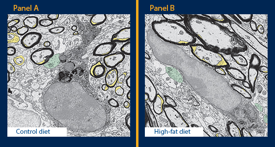
These pictures, taken by a scanning electron microscope, show the changes in the brain of offspring exposed to a normal diet (Panel A) vs a high-fat diet (Panel B) in utero. We can see the number of myelin channels (yellow) decreasing in the male offspring exposed to the high-fat diet in utero while their microglia (black) are making more contacts with synapses (presynaptic terminals in green and postsynaptic dendrites in red) – Photo by Fernando González Ibáñez.