Research highlights

Dr. Reuven Gordon and his group discover a new, inexpensive way to detect cancer. A new generation of sensors based on nano plasmonics are coming out soon.

Regie Sobral-Filho, a PhD student who works in Dr. Alex Brolo’s lab, is collaborating with scientists at the BC Cancer Agency to develop biologically adaptive radiotherapy.

Optical trapping specimen preparation: Abhay Kotnala (PhD student) is pictured preparing a specimen for optical trapping using the double nanohole on a 100 nm gold thin film.

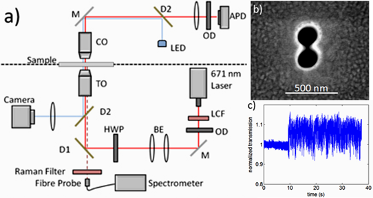
Steven Jones working on the optical trapping set-up. A laser tweezer system is used to study individual dielectric nanoparticles (~20nm, TiO2/Polystyrene) and identify them using SERS.
Radiation-induced glycogen accumulation detected by single cell raman spectroscopy is associated with radioresistance that can be reversed by metformin
Altered cellular metabolism is a hallmark of tumor cells and contributes to a host of properties associated with resistance to radiotherapy. Detection of radiation-induced biochemical changes can reveal unique metabolic pathways affecting radiosensitivity that may serve as attractive therapeutic targets.
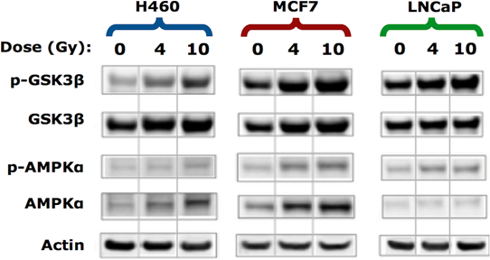
Using clinically relevant doses of radiation, we performed label-free single cell Raman spectroscopy on a series of human cancer cell lines and detected radiation-induced accumulation of intracellular glycogen. The increase in glycogen post-irradiation was highest in lung (H460) and breast (MCF7) tumor cells compared to prostate (LNCaP) tumor cells. In response to radiation, the appearance of this glycogen signature correlated with radiation resistance. Moreover, the buildup of glycogen was linked to the phosphorylation of GSK-3β, a canonical modulator of cell survival following radiation exposure and a key regulator of glycogen metabolism.
When MCF7 cells were irradiated in the presence of the anti-diabetic drug metformin, there was a significant decrease in the amount of radiation-induced glycogen.The suppression of glycogen by metformin following radiation was associated with increased radiosensitivity. In contrast to MCF7 cells, metformin had minimal effects on both the level of glycogen in H460 cells following radiation and radiosensitivity. Our data demonstrate a novel approach of spectral monitoring by Raman spectroscopy to assess changes in the levels of intracellular glycogen as a potential marker and resistance mechanism to radiation therapy. Read more.
Polarization-dependent extraordinary optical transmission from upconversion nanoparticles
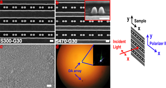
Enhanced upconversion (UC) emission was experimentally demonstrated using gold nanoparticles double antennas coupled to nanoslits in gold films. The transmitted red emission from UC ytterbium and erbium co-doped sodium yttrium fluoride (NaYF4:Yb3+/Er3+) nanoparticles (UC NPs) at ~665 nm (excited with a 980 nm diode laser) was enhanced relative to the green emission at ~550 nm. The relative enhanced UC NPs emission could be tuned by the different polarization-dependent extraordinary optical transmission modes coupled to the gold nanostructures.
Finite-difference time-domain calculations suggest that the preferential enhanced UC emission is related to a combination of different surface plasmon modes excitation coupling to cavity Fabry-Perot interactions. A maximum UC enhancement of 6-fold was measured for nanoslits arrays in the absence of the double antennas. In the presence of the double nanoantennas inside the nanoslits, the UC enhancement was between 2 and 4-fold, depending on the experimental conditions. Read more.
Nano particle application in radiotherapy
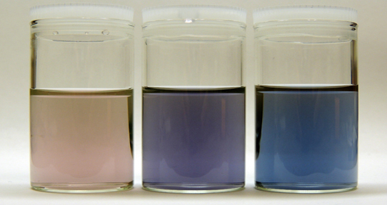
Nano particles that are 10,000 times thinner than a human hair. Core-shell nano structures have the advantage of combining two or more properties from different types of materials, generating features that can be highly dependent on the size of the nanostructures.
Picture bellow shows that the optical properties of silver nanoshells can be tuned by different core sizes. Regie Sobral-Filho, a PhD student who works in Dr. Alex Brolo’s lab, is collaborating with scientists at the BC Cancer Agency to develop biologically adaptive radiotherapy, treatment planning based on a patient’s individual biochemistry. He also uses inexpensive and accessible nano fabrication techniques so that his methods can be replicated in labs all over the world.
Raman spectroscopy of single nanoparticles in a double-nanohole optical tweezer system
A double nanohole in a metal film was used to trap nanoparticles (20 nm diameter) and simultaneously record their Raman spectrum using the trapping laser as the excitation source. This allowed for the identification of characteristic Stokes lines for titania and polystyrene nanoparticles, showing the capability for material identification of nanoparticles once trapped. Increased Raman signal was observed for the trapping of multiple nanoparticles. This system combines the benefits of nanoparticle isolation and manipulation with unique identification. Read more.
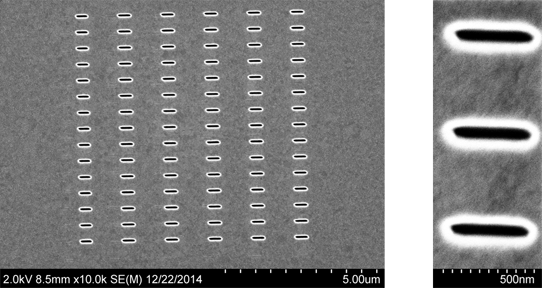
Localized and propagating surface plasmon resonances in aperture-based third harmonic generation
Scanning electron microscopy (SEM) images of an array of rectangular apertures fabricated using focussed ion beam milling on a 100 nm gold film. These structures were designed to study the origin of third harmonic generation from metasurfaces. By Mohammadreza Sanadgol Nezami from Dr. Reuven Gordon's group.